Intraoral Massage Certification
Here are some of the images for Intraoral Massage Certification that we found in our website database.

Intraoral Massage PDF Neck Massage

Intraoral Massage Certification prosecution2012

Intraoral Massage Certification prosecution2012

Massage Therapy Certification How to Elevate your Career

International Massage Therapy Course Certification and Accreditation

PPT SFH ORTHOPEDIC MASSAGE CERTIFICATION PROGRAM PowerPoint

Equine Massage Therapy Certification Course

National Certification Board for Therapeutic Massage and Bodywork on

Buccal Massage Certification prntbl concejomunicipaldechinu gov co

Buccal Massage Certification prntbl concejomunicipaldechinu gov co

Buccal Massage Certification prntbl concejomunicipaldechinu gov co

National Certification Board for Therapeutic Massage and Bodywork on

Neuromuscular Massage Certification prntbl concejomunicipaldechinu gov co

Intraoral Massage Certification Endorsement Elite Massage School

Intraoral Massage Certification Endorsement Elite Massage School

Intraoral Massage Certification Endorsement Elite Massage School

Neuromuscular Massage Therapy Certification prntbl

Kimberley Swanson Massage Therapist in Seattle WA

Lymphatic Drainage Massage Certification For Nurses prntbl

Certification vs Licensure in Esthetics Massage Therapy and
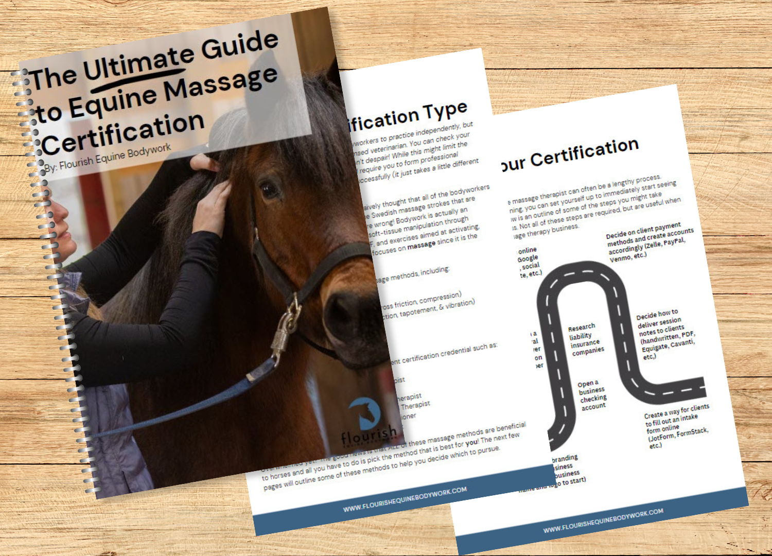.png "Ultimate Guide to Equine Massage Certification Digital Download")
Ultimate Guide to Equine Massage Certification Digital Download

Massage therapists are this and so much more 🙌 National

Indian Head Massage Certification Course Sunshine Coast

Intraoral Massage Self Transformations Massage

Massage Therapy Butter Massage Spa

Intraoral Massage Self Transformations Massage

Intraoral Massage Self Transformations Massage
About Kaarin Stone Lake Massage

Intraoral Massage Kenzie Renkert

Buccal Massage Discover this transformative power

How Can You Get Abhyanga Massage Training? Certification Explained
Intraoral Massage Absolute Body Balance

Salem Massage and Lymphatics Salem OR

PPT Massage Therapist Certification in Canada Get Certified Start

Ultimate Intraoral Scanning Certification E ssential Networks

Medical Massage Courses Certification Science of Massage Institute

How to Renew BIS Certification Complete Guide

Complete Guide to Smoking Laws in Thailand

Esalen® Massage Certification course BODY UNIVERSAL

Buccal Face Massage Vancouver RMT Intraoral Massage for TMJ Relief

Intraoral Massage: Things You Need to Know Vibrant Skin Bar

Buccal Massage Jaw Massage and De Armouring Intraoral Massage

45 Intraoral Massage Royalty Free Images Stock Photos Pictures

An Intraoral TMJ Massage Finally Relived My Jaw Pain

Taipei Massage Recommendation Guide》Mysterious Customer Certification

TMJ Intraoral Massage in Seattle

Wa Intraoral Massage Form Fill and Sign Printable Template Online

Does Medicaid Pay For Massage Therapy at Jennifer Reynolds blog

How To Do Intraoral Massage

Amazon com: Nicpower Massage Gun Charger Fit for Therabody Theragun

Physiotherapist Applying Intraoral Massage Therapy On Patient Stock

2024 Education Recap and What to Expect in the Future

Massage Virgin Coconut Oil Machine Certification : ISO9001:2008 at Rs

Intraoral Massage Therapy YouTube

ASMR Sculptural Face Lift and Intraoral Massage relaxldn

Morningmassage part two Buccal/intraoral massage to help with

Treatment of sialadenitis MEDizzy

Wenatchee Valley Massage Wenatchee Valley Massage Clinic Facebook

Bank 8 hours of CE Ohio Dental Hygienists #39 Association Facebook